

Precision Pain Care
Interventional Pain Management
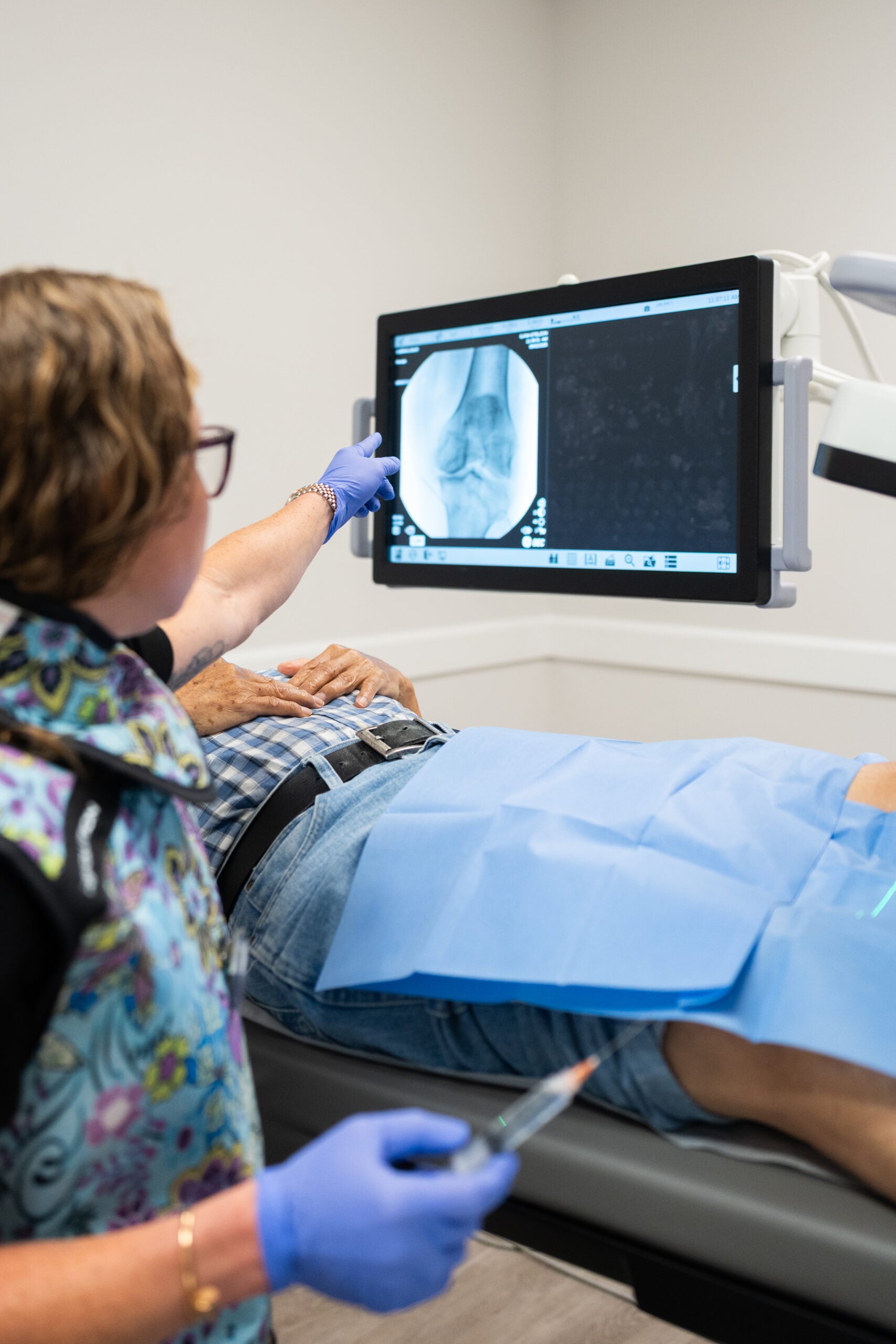
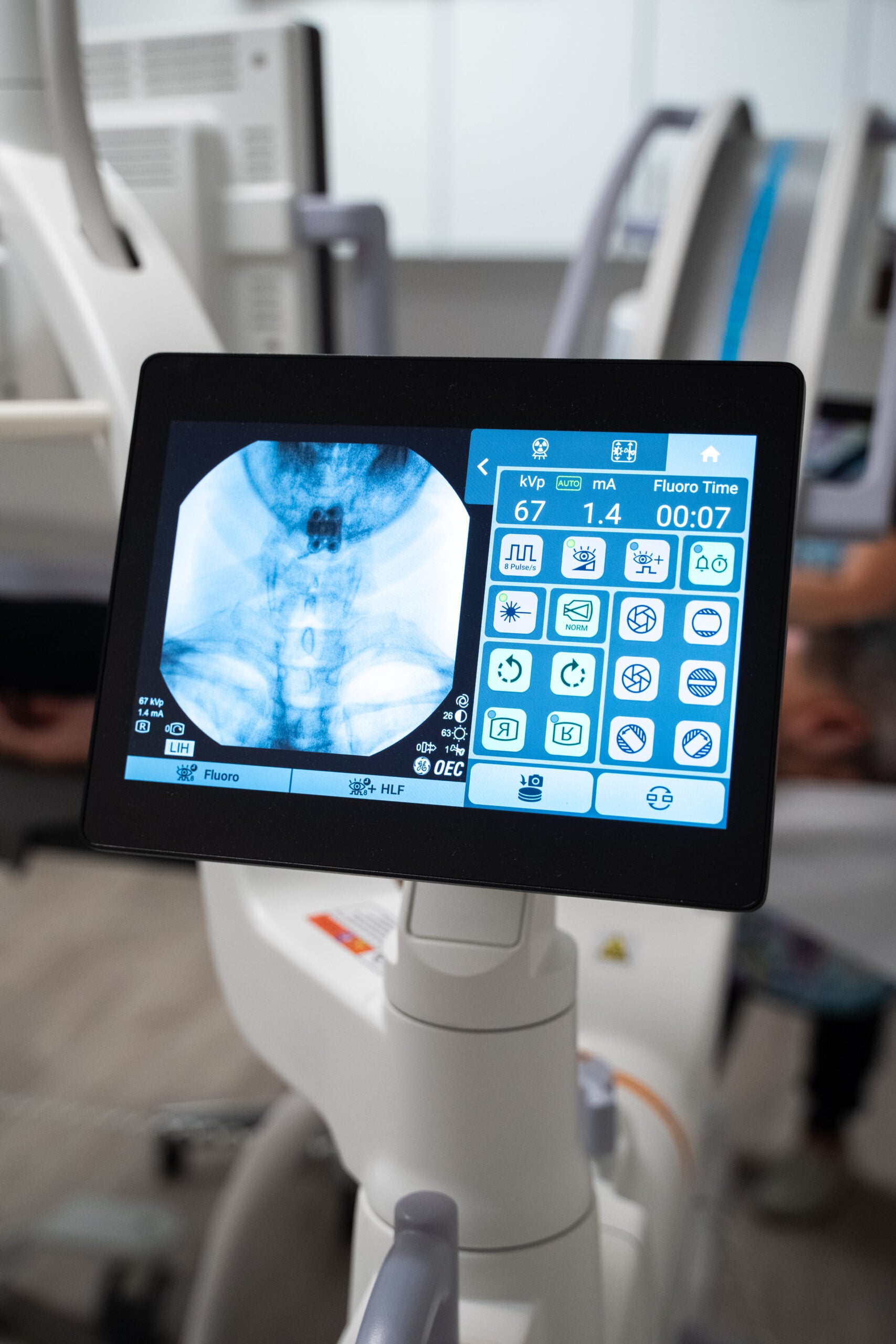
Every injection and minimally invasive procedure we perform, organized by what it treats. All performed under fluoroscopic or ultrasound guidance so placement is precise.
Diagnosis First, Then the Procedure
Pain rarely comes from where you feel it. Before anything is injected, Dr. Etelzon works to identify the actual structure generating your symptoms using examination, imaging review, and where appropriate diagnostic blocks.
That matters because the procedures below are precise tools. Placed into the right structure they can be transformative. Placed into the wrong one they do very little, which is the most common reason patients tell us a previous injection "did not work."
Many of these procedures pair well with regenerative treatments, and Dr. Etelzon will tell you when a biologic approach is the better long-term answer.
Category One
Spine Injections
Targeted injections into the specific spinal structure causing your neck or back pain, guided by fluoroscopy.
Epidural Steroid Injections
Placed into the epidural space to reduce inflammation around irritated nerve roots. Performed at cervical, thoracic, and lumbar levels depending on where the compression sits.
CervicalThoracicLumbarMedial Branch Blocks
A diagnostic block of the small nerves carrying pain signals from the facet joints. Used to confirm the facet joints are the source before proceeding to a longer-lasting treatment.
DiagnosticFacet Joint Injections
Delivered into the small paired joints at each spinal level. These joints become arthritic with age and are a frequent source of axial neck and back pain that worsens with extension.
Sacroiliac Joint Injections
Into the joint where the spine meets the pelvis. A commonly missed source of low back and buttock pain, particularly pain that is worse on one side when sitting or standing from a chair.
Category Two
Joint and Nerve Procedures
For pain originating in a peripheral joint, a specific nerve, or muscle tissue rather than the spine.
Joint Injections
Image-guided injections into any joint including knee, shoulder, hip, ankle, wrist, elbow, and the small joints of the hand and foot. Guidance matters here, since blind injections frequently miss the joint space entirely.
Peripheral Nerve Blocks
Targeted at a specific peripheral nerve to interrupt pain signaling. Used both diagnostically to confirm a nerve is involved and therapeutically to provide relief.
Trigger Point Injections
Into taut bands of muscle that refer pain elsewhere in the body. Useful for myofascial pain that has not released with manual therapy or stretching.
Category Three
Minimally Invasive Spine Procedures
Procedures that achieve surgical goals through small access points, generally with shorter recovery than open spine surgery.
Percutaneous Discectomy
Removal of disc material through a needle-sized access point to relieve pressure on a compressed nerve. Performed at both cervical and lumbar levels.
CervicalLumbarEndoscopic Lumbar Discectomy
Uses a small camera to visualize and remove herniated disc material directly, through an incision considerably smaller than traditional open discectomy.
Endoscopic Rhizotomy
Endoscopic visualization of the medial branch nerves for a more complete and durable interruption of facet-mediated pain than a percutaneous approach alone.
Kyphoplasty
Stabilizes a vertebral compression fracture by restoring height and injecting bone cement, often producing rapid relief of fracture pain.
MILD Procedure
Minimally Invasive Lumbar Decompression. Removes a small portion of thickened ligament narrowing the spinal canal, for patients with lumbar spinal stenosis whose main symptom is pain when walking or standing.
Peripheral Nerve Stimulation
PNS places a small lead near a specific peripheral nerve to modulate pain signals. A trial period is performed first so you know whether it works for you before anything permanent is placed.
Category Four
Radiofrequency Ablation
When a diagnostic block confirms the facet joints are the source of your pain, radiofrequency ablation offers considerably longer relief than an injection alone. It uses heat generated by radio waves to interrupt the small medial branch nerves carrying pain signals from those joints.
Because those nerves only carry sensation and do not control movement or strength, interrupting them relieves pain without affecting function. Relief commonly lasts six to twelve months or longer, and the procedure can be repeated if nerves regenerate.
Pain Doctors Medical performs radiofrequency ablation using Stryker systems.
Two approaches
Continuous Radiofrequency
Sustained heat creates a precise lesion on the target nerve, interrupting pain transmission. The standard approach for confirmed facet-mediated neck and back pain.
Pulsed Radiofrequency
Delivers energy in pulses at lower temperature, modulating the nerve rather than creating a lesion. Used where a continuous lesion would not be appropriate, including around certain nerves where preserving the nerve matters.
Who it helps most
Patients with confirmed facet joint pain in the neck or lower back, particularly those who got good but temporary relief from a medial branch block. It is also used for sacroiliac joint pain and, in selected cases, for knee pain through genicular nerve ablation.
Category Five
Surgical Options
When conservative and minimally invasive approaches are not enough, Dr. Alexandra Carrer provides surgical evaluation and treatment within the same practice, so you are not starting over with a new team.
Arthroscopy
Small-incision joint surgery using a camera to diagnose and treat problems inside the joint, most commonly the knee and shoulder.
Lumbar Discectomy
Surgical removal of herniated disc material compressing a nerve root, for patients with persistent radicular pain or neurologic findings that have not resolved with other treatment.
Minimally Invasive Spinal Surgery
Spinal procedures performed through smaller incisions with less muscle disruption than traditional open surgery, generally allowing faster recovery.
Surgery is a decision, not a default
Dr. Etelzon's practice is built on non-operative treatment, and most patients never need this section. When imaging shows a full-thickness tear, or an examination reveals progressive weakness, significant numbness, or loss of function, prompt surgical consultation is the right answer and you will be told so directly.
Your Visit
What to Expect
- BeforeYour imaging and history are reviewed, and the specific structure being targeted is explained to you before anything is scheduled.
- Image guidanceProcedures are performed under fluoroscopy or ultrasound. You will be positioned comfortably and the skin is numbed first.
- DurationMost injections take a few minutes. Minimally invasive procedures take longer but are still same-day, outpatient, and typically under local anesthetic with sedation as needed.
- AfterwardSome soreness at the site is normal for a day or two. Specific activity guidance depends on the procedure and will be given to you in writing.
- InsuranceMost procedures in this category are covered by Medicare and commercial insurance, unlike regenerative treatments. Our team verifies coverage before scheduling.
"An injection that goes to the wrong structure is not a failed treatment. It is a failed diagnosis. Getting that part right is most of the work."
Dr. Ilana Etelzon, MD